
Laboratory and Diagnostic Imaging

Our in-house diagnostic laboratory enables us to perform a variety of routine diagnostic procedures such as blood tests, analysis of urine, fecal analysis, cytology (examining tissue samples) and heartworm testing. Our in-house imaging (endoscope, digital x-ray, dental digital x-ray and ultrasound) allows us to do further diagnosis without any invasive surgery on your pet.

Ultrasound

By

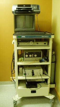
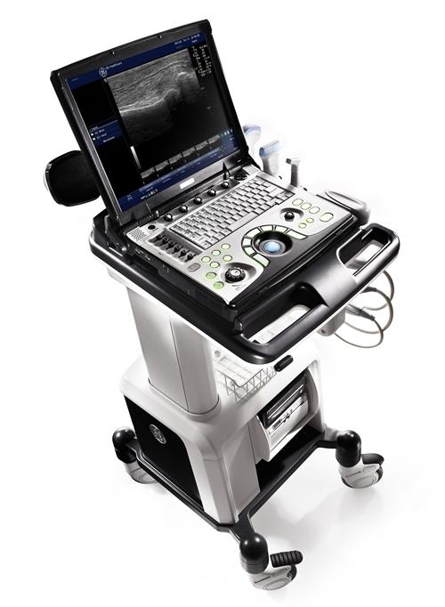
Veterinary ultrasound is a useful tool which can be used to diagnose and monitor a variety of conditions. In some circumstances, it may be necessary to use veterinary ultrasound because animals cannot communicate with humans about their symptoms. As a result, an ultrasound exam may be used to asses a pet for otherwise unseen or undetected problems.

One of the most common reasons for the doctor to use ultrasound is as an aid to diagnosis. For example, if a pet is brought in by an owner who claims that the animal is having difficulty urinating, the doctor might use ultrasound to check for an obstruction in the bladder or urethra. Ultrasound can also be used to examine suspicious masses and other abnormalities which are detected during a physical exam. It can also be used in emergency settings to look for serious medical problems such as internal organ damage after a collision.

We can also use veterinary ultrasound to monitor an ongoing condition. A pregnant dog, for example, may be given several ultrasound examinations to confirm the pregnancy and assess the health of the developing puppies. Ultrasound can also be used to monitor the progress of liver and kidney disease, along with any treatment approaches, just as it is in humans.

Depending on the findings of the ultrasound study, the doctor can recommend the best course of action, which can vary from requesting additional testing or procedures to recommending a change of medication to continuing the animal’s care as before.

Digital Dental Radiographs (X-rays)

By

Periodontal disease is the most common ailment in companion animals. Dental radiographs can reveal significantly more problems than a thorough oral exam.

Patients in for “routine teeth cleaning and examination” will now benefit from a doctor seeing the other 50% of the tooth below the gum line. This allows the doctor to detect serious issues that would be impossible to diagnose without radiographs. Some of these issues are enamel defects, fractures beneath the gum line and bone loss.

Radiographs permanently document the problem in black and white. The degree of bone loss can be measured. Clients can see the areas of bone loss around the tooth, or a resorption that has “eaten away” the tooth’s root. Also, the radiograph can be compared to “normals” so clients can easily see an abnormal result.

The digital machine allows the doctors to use less radiation and has a more rapid turnaround. Also, they are then able to manipulate the images on the screen and adjust as required.

The dental radiograph becomes part of your patient’s permanent medical record. Examining serial radiographs of periodontal or endodontic cases taken at three to six-month intervals provides invaluable information concerning progression or resolution of disease.

Laboratory

By

Our in-house diagnostic laboratory enables us to perform a variety of routine diagnostic procedures such as blood tests, analysis of urine, fecal analysis, cytology (examining tissue samples) and heartworm testing.

The ability of our staff to perform these tests quickly and accurately in clinic enables us to provide a timely and accurate diagnosis of your pet’s condition, and allows us to start appropriate treatment of that condition as quickly as possible. It also allows us to perform pre-anesthetic blood testing on your pet immediately prior to any needed surgical procedure, making both the procedure and the necessary anesthetic safer for your pet. In addition, we can perform wellness screening (recommended annually for any pet over seven years of age) which can lead to the detection of hidden health problems and allow for early treatment.

In some cases, it is necessary to enlist the help of outside laboratories or diagnostic specialists in order to properly diagnose your pet’s health problems.

Endoscopy

By

An endoscope is a medical instrument used for visual examination of the interior of a body cavity or a hollow organ such as the colon, bladder, or stomach, and for laparoscopic surgery. It is a rigid or flexible tube fitted with lenses (like a camera), a fiber-optic light source, and often a probe, forceps, suction device, or other apparatus for examination or retrieval of tissue.

Endoscopy is a wonderful diagnostic tool. Essentially, endoscopic equipment allows us to look inside the body in a far less invasive manner than traditional surgical techniques. At our clinic, we use endoscopy most commonly to check the esophagus and stomach for obstructions or foreign bodies. We can also search for the cause(s) of chronic vomiting and diarrhea by looking in the stomach, colon or esophagus.

A routine ear examination using a hand-held otoscope could miss easily treatable problems. By using our in-clinic video otoscope, we are able to dramatically improve our visualization of the ear canal and provide better and more accurate treatment. Because it records images at a magnification of 15 times, we can even see ear mites right on the video screen!

Often we will perform biopsies (for example: liver, bowel, kidney) using endoscopic equipment because it is far less invasive than traditional surgical methods. We can even perform bladder stone removal endoscopically in many cases.

The main advantages of endoscopy are as follows:

  • allows the veterinarian to look inside body cavities to search for disease
  • allows for minimally invasive procedures resulting in quicker recoveries and fewer complications
  • better diagnostics equals more accurate and better treatments = better pet health
  • smaller incisions
  • may be used for all animals including avians and exotics

To find out if endoscopy is right for your pet, speak with our staff and learn more about this innovative technology.

Digital Radiology (X-rays)

By

When we need to figure out what’s wrong with your pet, we routinely use x-rays to help identify the cause of the problem, rule out possible problems, or provide a list of possible causes. We may also use x-rays during a wellness exam to diagnose potential problems before they become serious.

X-rays provide valuable information about a pet’s bones, gastrointestinal tract (stomach, intestines, colon), respiratory tract (lungs), heart, and genitourinary system (bladder, prostate). We use radiology alone or in conjunction with other diagnostic tools. Interpretation of radiographs requires great skill on the part of the veterinarian.

We offer digital radiology (x-rays that are captured digitally rather than on film). This technology allows us to provide you with a quicker diagnosis for your pet. Plus, it uses less radiation than traditional x-rays.

To avoid a blurry image, pets need to remain completely still while an x-ray is taken. In some cases, we may need to sedate your pet or use short-acting general anesthesia.

If you have any questions about our radiology service or what to expect during your pet’s procedure, please don’t hesitate to ask.

We're accredited by the American Animal Hospital Association